
By Dr Margi Hirapara
Consultant Breast Surgeon
July 05, 2025
Breast cancer remains the most common cancer affecting women across the world. In India, the incidence of breast cancer is steadily rising, and early detection is vital for successful treatment and improved survival. Approximately 99% of breast cancers occur in women, and 0.5–1% of breast cancers occur in men. (Source:https://www.who.int/news-room/fact-sheets/detail/breast-cancer) With these numbers, screening plays a critical role.
At KD Hospital, we are proud to house Gujarat’s leading team of medical, surgical, and radiation oncologists under one roof. Our team uses a comprehensive, multidisciplinary approach to cancer care by integrating advanced diagnostic tools, innovative surgical techniques such as robotic-assisted surgery, and personalised treatment plans.
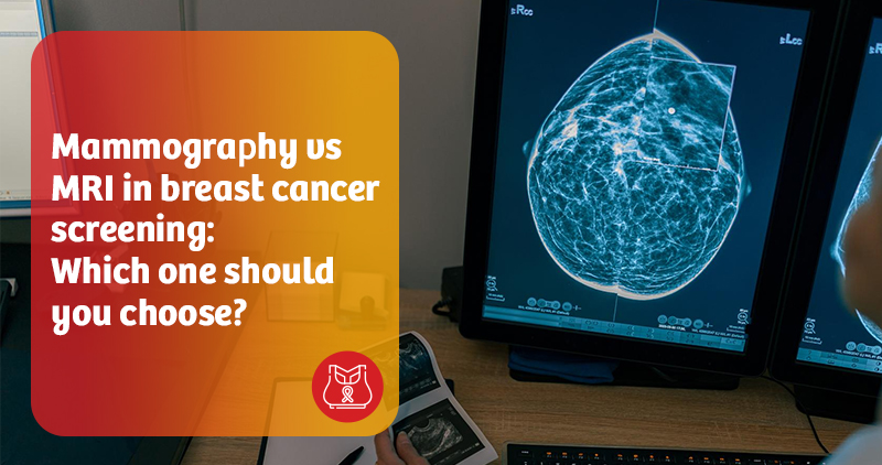
When it comes to breast cancer screening, mammography and breast MRI (Magnetic Resonance Imaging) are the two most commonly discussed options. But the question remains—which is right for you? Let’s break down the science, strengths, and ideal use.
Mammography is the most widely used screening tool for breast cancer. It involves a low-dose X-ray that can detect changes in breast tissue, including small tumours that may not be prominent.
Breast MRI uses magnetic fields and radio waves to create detailed cross-sectional images of breast tissue. MRI, unlike mammography, does not involve radiation and offers high sensitivity in detecting even subtle abnormalities.
Breasts are composed of glandular, fibrous, and fatty tissues. Women with more glandular and fibrous tissues are said to have dense breasts. On a mammogram, dense tissue appears white—the same as many tumours—making it harder to detect cancer.
Note: Dense breasts are common in younger women, especially those under the age of 50. Your doctor may recommend additional imaging based on your breast density.
| Feature | Mammography | MRI |
| Technology | X-ray | Magnetic waves |
| Radiation | Yes | No |
| Sensitivity | Moderate | High |
| Specificity | High | Moderate |
| Best for | Average-risk women | High-risk women |
| Contrast dye | Not needed | Required |
| Time | 10–15 minutes | 30–60 minutes |
For average-risk women
For high-risk women
High risk is defined as a lifetime breast cancer risk of 20–25% or higher. It includes
Pros and cons at a glance
Advantages
Limitations
Advantages
Limitations
In some cases, combining both tests offers superior diagnostic value. Each modality has unique strengths.
MRI is excellent at picking up invasive cancers, especially in dense tissue.
Mammography is better at detecting calcifications, which can signal ductal carcinoma in situ (DCIS)—an early form of breast cancer.
Combined screening improves the chances of catching cancer early when it is most treatable.
At KD Hospital, we prioritise early detection, precise diagnosis, and personalised care for all our patients. Our breast cancer screening protocols are tailored based on individual risk assessments, genetic profiles, and breast density.
Best hospital in Gujarat for breast cancer treatment.
The choice between mammography and MRI depends on your risk factors, age, and breast density.
For most women at average risk, mammography remains the gold standard for routine screening. For individuals at higher risk, an MRI can provide crucial insights that complement mammography and potentially save lives.
Before making a decision, consult a breast specialist or oncologist. A personalised screening strategy can make all the difference in early detection and successful treatment.
